


")


EVIS X1™ Video System Center


Video System
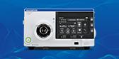
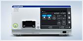
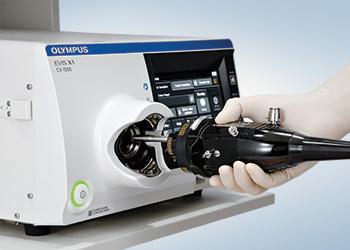
EVIS X1™ Video System Center
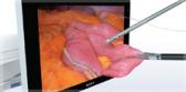
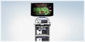
The EVIS X1 CV-1500 Video System Center combines video processor and LED light source in one box, resulting in a compact and lightweight design.1 The CV-1500 features multiple special observation modes that support physicians in every step during endoscopic procedures.
Key Benefits
In addition to conventional white light and NBI™ (Narrow Band Imaging™) Technology, the CV-1500 offers three other powerful enhanced observations to improve diagnostic and therapeutic capability:
- TXI™ Technology (Texture & Color Enhancement Imaging) optimizes the structure, color tone and brightness of the mucosal surface.2
- RDI™ Technology (Red Dichromatic Imaging) improves visibility of deep blood vessels and bleeding points.2
- BAI-MAC™ (Brightness Adjustment Imaging with MAintenance of Contrast) technology improves brightness in darker portions of the image.2
TXI, RDI, NBI and BAI-MAC technologies are not intended to replace histopathological sampling as a means of diagnosis.
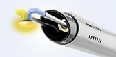
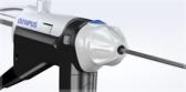
With One-Touch Connector and no need for white balance adjustment,3 setup is simplified, with the aim of streamlining workflow and accelerating procedure time. Touch-sensitive panel facilitates instinctive operation, while convenient functions like Pre-freeze and MyCV mode assist in creating a user-friendly working environment. Downtime may be reduced thanks to the use of LED bulbs that have been designed to last years without needing replacement.4
1. Data on file with Olympus (DC00436067)
2. Data on file with Olympus (DC00489968)
3. Olympus 1100 series endoscopes only, data on file with Olympus (DC00436067)
4. Data on file with Olympus (DC00468069)
Complications from an endoscopic procedure may include, but are not limited to, reaction to sedation, bleeding, perforation, infection, and/or pancreatitis. Never apply the endoscope connected to the CV-1500 video system center to the heart or any area near the heart; leakage current from this Type BF-applied part may be dangerous and cause ventricular fibrillation or otherwise seriously affect the cardiac function of the patient. Improper disinfection of the video system center may result in operator and/or patient infection. For complete indications, contraindications, warnings and cautions, please reference the Instructions for Use (IFU) that accompanied the EVIS X1™ CV-1500 video system center.
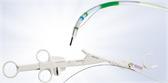
Related Products

Product Support
Power Supply |
Rated voltage |
100-240 V AC; Within ±10% |
Frequency |
50/60 Hz; within ±3 Hz |
|
Rated input |
600 VA |
|
Size |
Dimension (W x H x D) |
370 x 198 x 488 mm 398 x 218 x 580 mm (maximum) |
Weight |
19.4 kg |
|
Classification (Medical Electrical Equipment) |
Type of protection against electric shock |
Class I |
Degree of protection against electric shock of applied part |
Depend on applied part. (The degree of protection against electric shock of this product is BF type if the mounting part to be connected to this product is BF type. However CF type is not subject to combination in this product.) |
|
Degree or protection against explosion |
The video system center should be kept away from flammable gases. |
|
Observation |
Analog signal output |
VBS composite |
Digital signal output |
12G-SDI 3G-SDI HD-SDI SD-SDI |
|
User settings |
The function settings for up to 20 users can be stored. |
|
Color tone adjustment |
Adjust the color tone of each endoscopic image for White light observation mode, NBI observation mode, and RDI observation mode. Red adjustment : ±8 steps · Blue adjustment : ±8 steps · Chroma adjustment : ±8 steps |
|
Automatic gain control (AGC) |
The image can be electronically amplified when the light is inadequate due to the distal end of the endoscope being too far from the object. |
|
Contrast |
H (High): Darkens the dark part and brightens the bright part. L (Low): Brightens the dark part and darkens the bright part. |
|
BAI-MAC™ Technology |
Brightness adjustment with maintenance of contrast |
|
Iris |
The iris modes can be switched. · Auto: The brightness is adjusted based on the brightest part of the central part and the average brightness of the periphery part. · Peak: The brightness is adjusted based on the brightest part of the endoscopic image. Average: The brightness is adjusted based on the average brightness of the endoscopic image. |
|
Image enhancement settings |
Fine patterns or edges in the endoscopic images can be enhanced electrically to increase the image sharpnes. |
|
Switching the enhancement modes |
The enhancement level can be selected from 3 levels (OFF, 1, 2, and 3) |
|
Image size selection |
The size of the endoscopic image can be selected from 2 modes. (Except SDTV) |
|
Electric zoom |
Switch between mode 1, mode 2, and mode 3. |
|
PIP/POP |
Switch between PIP and POP. |
|
Aspect ratio |
Switch between 16:9 and 4:3. (Except SDTV) |
|
Freeze |
Freeze the endoscopic image. |
|
Pre-freeze |
The image with the least blur is selected from the images captured in the set time period before freeze operation and displayed. |
|
Optical-digital observation |
The optical-digital observation can be performed with compatible endoscope.
| |
Custom switch |
Assign specific functions to the following buttons. · Remote switches (Up to 5) · Foot switches (Up to 2) · Keyboard custom key (Up to 4) · Touch panel custom button of basic functions screen (Up to 3) · Touch panel custom button of custom functions screen (Up to 10) |
|
MyCV mode |
Customizable switch to activate multiple functions at once. |
|
Documentation |
Remote control |
The following peripheral device can be controlled (specified models only). |
Patient information |
The following data can be displayed on the monitor. · Patient ID · Patient name · Gender · Age · Date of birth · Comment |
|
Displaying the recording state |
The recording state of the following peripheral device can be displayed on the monitor. · Portable memory: Remaining capacity |
|
Displaying the image information |
The following data can be displayed on the monitor. · Image enhancement · Electric zoom ratio · Color mode · Focus · Observation mode |
|
Advanced registration of patient information |
Up to 50 patients information can be registered. · Patient ID · Patient name · Gender · Age · Date of birth |
|
Recording format |
Standard image quality: TIFF; Low image quality: JPEG |
|
Memory Backup |
Memorization of user settings |
The settings are retained when the video system center is turned OFF. |
White balance |
Once the white balance is set for a specific scope it is held in the systems memory. |
Olympus® Service & Repair
Olympus offers a broad range of services to healthcare professionals and to our customers, including contact hour and peer-based training courses; information, training tools and videos on infection control and reprocessing; authorized repair services and support on Olympus equipment; and financing solutions to help your facility with acquisition of new capital equipment, accessories, and maintenance plans.