


")


Texture and Color Enhancement Imaging
TXI™ Technology – See Things in a New Light
One of several innovations available with the EVIS X1™ endoscopy system, Texture and Color Enhancement Imaging (TXI™) technology was designed to increase the visibility of potentially suspicious lesions and polyps by enhancing image color and texture during endoscopic screening.1
Early detection is critical for cancer prevention and decreasing mortality.2 A 1% increase in the adenoma detection rate (ADR) results in a 3% decrease in the risk of interval cancer and a 5% decrease in the risk of fatal interval colorectal cancer. However, precursor lesions are often tiny and easy to overlook, so good visibility is essential in characterizing and treating these lesions more effectively.2,3
A multicenter randomized controlled trial published in Gastroenterology in October 2023 revealed TXI technology significantly improves the ADR by 13.61%, as well as the rate of adenomas per colonoscopy (APC) ≥ 5mm in size, versus white light endoscopy (WLE), highlighting its ability to support clinicians in identifying potential precancerous lesions and enhancing the quality of their colonoscopies.4
How It Works
TXI technology is designed to emphasize image information by combining the three image processing algorithms: brightness correction of the dark part of the image; color difference expansion processing; and texture component emphasis processing.1
The incoming image is split, and the texture and brightness are enhanced before the separate images are merged back together. Additional color enhancements are made to define subtle tissue differences more clearly.5
TXI technology is applied during post-processing; therefore, the color appearance of the final image is like white light, making the viewing of lesions more relatable for the user.5
The following video explains how TXI technology works and its purpose during an endoscopic procedure.
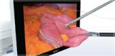
The following video shows Prof. Dr. Jürgen Pohl using an EVIS X1 GIF-1100 gastroscope to observe a duodenal adenoma under TXI technology.
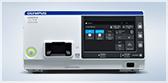
Learn More About The EVIS X1™ Endoscopy System
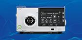
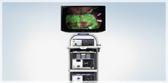
Learn More about the CV-1500 Video System Center
Learn More about BAI-MAC™ Technology
1. Data on file with Olympus (DC00489968).
2. American Cancer Society: Colorectal Cancer Facts & Figures 2017-2019, p. 15. https://www.cancer.org/content/dam/cancer-org/research/cancer-facts-and-statistics/colorectal-cancer-facts-and-figures/colorectal-cancer-facts-and-figures-2017-2019.pdf.
3. Corley DA, Jensen CD, Marks AR, et al. Adenoma detection rate and risk of colorectal cancer and death. N Engl J Med. 2014;370(14):1298-1306.
4. Young E, Rajagopalan A, Tee D, et al. Texture and color enhancement imaging improves colonic adenoma detection:
A multicenter randomized controlled trial. Gastroenterology. 2024;166(2):338-340.e3.
5. Data on file with Olympus (DC00785702).
The EVIS X1 endoscopy system is not designed for cardiac applications. Other combinations of equipment may cause ventricular fibrillation or seriously affect the cardiac function of the patient. Improper use of endoscopes may result in patient injury infection, bleeding, and/or perforation. Complete indications, contraindications, warnings, and cautions are available in the Instructions for Use (IFU).
TXI technology is not intended to replace histopathological sampling as a means of diagnosis.
TXI is a trademark of Olympus Corporation, Olympus America, Inc., and/or their affiliates.
Clinical video provided by Prof. Dr. Jürgen Pohl using a GIF-1100 gastroscope.