Endoscopic Ultrasound (EUS)
Commited to
Definitive
Diagnosis
Endoscopic Ultrasound
Diagnose and treat with confidence.
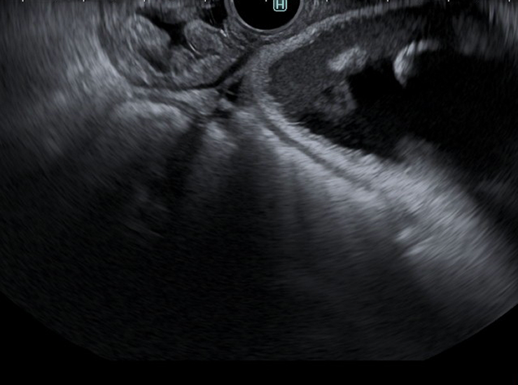
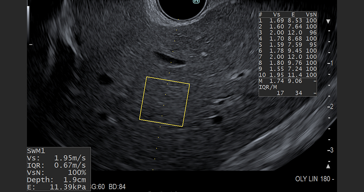
Endoscopic ultrasound (EUS) combines ultrasound and conventional endoscopy technologies to see beyond the walls of the gastrointestinal tract. EUS allows endoscopists to visualize all five layers of the GI tract, as well as surrounding tissue and organs.
Olympus offers a full product portfolio of EUS processors, echoendoscopes, miniature probes and needles to meet physician needs and preferences. These products can support diagnostic, therapeutic and interventional applications such as EUS-guided tissue acquisition, cyst drainage and biopsies of lesions/lymph nodes.
Access
The Olympus® portfolio offers a variety of echoendoscopes and miniature probes, creating a total endosonography solution for a full range of minimally invasive applications.
The diagnostic radial array echoendoscope provides a 360°, cross-sectional view of the GI tract and is primarily used for screening. The curvilinear echoendoscopes are useful for therapeutic applications such as tissue sample collection, cyst drainage, biopsies of lesions/lymph nodes and injection for pain management.
In addition, the endoscopic ultrasound miniature probes generate 360° images in the near field for visualization of submucosal areas.
Visualization
Visualization is key to accurately identifying tumors and tissue properties and boundaries. Advanced features available on Olympus® ultrasound processors are designed to improve visualization and assist in diagnostic, therapeutic, and interventional procedures, that may lead to improved quality of patient care.
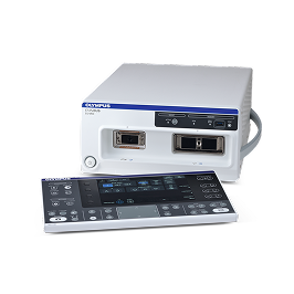
Olympus offers a full line-up of ultrasound processors designed to meet and exceed the needs of gastroenterologists performing a wide range of EUS procedures.
Therapeutic EUS
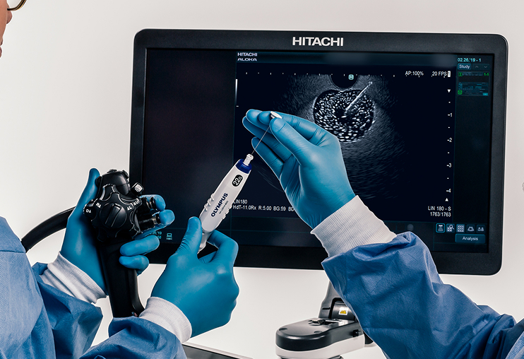
Olympus offers a line-up of single-use FNB and FNA needles to help achieve procedural goals for endoscopic ultrasound in a variety of clinical settings.
The EZ Shot™ 3 Plus FNB needle offers access to lesions in difficult locations, enhanced echogenicity, and reduced puncture force in 19 G, 22 G, and 25 G configurations.1
The EZ Shot 3 FNA needle features the same ergonomic handle as the EZ Shot 3 Plus needle and is designed for smooth puncturability of even small target lesions, available in 19 G, 22 G and 25 G configurations to suit clinical preferences.2
EUS Procedure Risks:
Potential complications that may be associated with endoscopic ultrasound include, but are not limited to, the following: sore throat, infection, bleeding, perforation, and/or tumor seeding (when EUS-FNA or FNB is performed). Do not use contrast agents when using the shear wave function. Do not use the shear wave function during puncturing or the interposition of any other type of metal. Contrast imaging and shear wave elastography functions are for GI only.
Products
At Olympus, we are committed to innovation. We deliver today’s most comprehensive EUS solution, with a robust portfolio of EUS processors, echoendoscopes and devices.
Endoscopic Ultrasound is the difference between maybe and definitely.
ALOKA ARIETTA™ 850
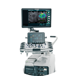
Premium ultrasound processor provides endoscopists with a depth of information allowing for decisive decision making, partnered with service and support. “Image quality” and “advanced technology” are two key areas where a determined effort has been made to refine fundamental performance and examination precision.
EU-ME3
High-quality compact ultrasound processor for use with Olympus® endoscopic and endobronchial ultrasound (EUS and EBUS) equipment that has been designed for integration with conventional endoscopy on a single workstation. With its high-resolution imaging and a display that promotes clear visualization, the EU-ME3 ultrasound processor brings real clarity to your EUS and EBUS procedures, supporting detection and characterization of lesions.
Optional features such as shear wave elastography, strain elastography and contrast harmonic imaging are available for the EU-ME3 and ARIETTA 850 ultrasound processors. Click on each of the product links to learn more.
EUS Processor Risks:
High output and prolonged exposure to ultrasonic waves can adversely affect the internal tissues of the patient. Scan only for the minimum length of time necessary for the diagnosis, and at the lowest possible output. Improper care, installation, or use, can cause electric shocks, burns or other injuries. The ultrasound endoscope connected to this ultrasound center must never be applied directly to the heart as it could cause ventricular fibrillation or otherwise seriously affect the cardiac function of the patient. Never allow an EndoTherapy accessory or another ultrasound endoscope, applied to or near the heart, to come in contact with the ultrasound endoscope connected to this ultrasound center. Do not use contrast agents when using the shear wave function, as there is a risk of injury to the patient’s tissue, resulting in bleeding, due to the interaction between the acoustic pressure of the ultrasound and the contrast agent. Do not use the shear wave function during puncturing or the interposition of any other type of metal as it may cause problems during the procedure. Contrast imaging and shear wave elastography functions are for GI only.
Curved Linear Array Echoendoscopes
GF-UCT180
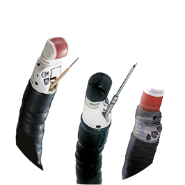
Curved linear array echoendoscope used for therapeutic applications such as tissue sample collection, cyst drainage, biopsies of lesions/lymph nodes and injection for pain management. The GF-UCT180’s ultrasound transducer has a broad bandwidth and high sensitivity, offering ultrasound imaging with high resolution and penetration and less noise when operating in B-mode.3
Radial Array Echoendoscopes
GF-UE160-AL5
Diagnostic radial array echoendoscope used to assess anatomy of the GI tract and surrounding structures. It provides a 360° cross-sectional view of the GI tract and is primarily used for screening.
EUS Scope Risks:
Complications from use of the GF-UCT180 or the GF-UE160-AL5 may include, but are not limited to: patient injury, bleeding, and/or perforation. Improper cleaning of an endoscope may result in patient infection. Balloons used with this instrument contain natural rubber latex that may cause allergic reactions. Do not use the balloon on a latex-sensitive patient.
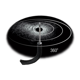
Olympus offers a full range of EUS and EBUS ultrasound miniature probes that can be used for various procedures. These miniature probes can be used for passing strictures, reaching peripheral lung lesions, conducting intraductal ultrasound and more.
Miniature Probes
Generate 360° high-resolution images in the near field for visualization of submucosal areas4.
Probe-driving Unit
Designed to support a spectrum of advanced EUS and EBUS procedures. The unit powers a wide variety of Olympus® ultrasound miniature probes with frequency ranges of 7.5 to 20 MHz.
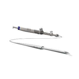
EZ Shot™ 3 Plus
FNB needle provides access to difficult to reach lesions without compromising scope position. The combination of the multi-layer coil sheath, nitinol needle*, and Menghini tip allow for precise and smooth penetration into the target lesion.1
EZ Shot 3
FNA needle offers enhanced visibility under ultrasound, with an anti-slip handle design that enables control when sampling.2
*nitinol material includes 19G and 22G only
EUS Needle Risks:
Complications from extra-luminal EUS guided FNA and FNB may include, but are not limited to infection, bleeding, perforation, and/or tumor seeding. Extra-luminal fine needle aspiration of cystic lesions has a higher risk of complication from infection and hemorrhage.
Endohepatology
Endoscopy + Hepatology
Traditionally, hepatologists refer liver patients to interventional radiology (IR) for liver imaging, biopsy, and management of portal hypertension.
Now, due to recent advancements in endoscopic ultrasound (EUS), liver imaging, shear wave measurement, contrast harmonic imaging, liver biopsy and portal pressure gradient measurement can be accomplished under EUS-guidance.
Thus, the integrated field has emerged as a result of the following5:
- Increased number of liver-focused endoscopic procedures
- Advances in endoscopic evaluation of the liver
- Advances in hepatology requiring liver assessment and histology

It would be most ideal if the assessment and treatment of liver disease and portal hypertension could be performed and assimilated by the primary liver/gastrointestinal (GI) specialist. We see this integration among specialists in esophageal and pancreaticobiliary diseases. It should be no different in hepatology.”
– Dr. Kenneth J. Chang
Gastroenterologist
Dr. Kenneth J. Chang is a consultant to the Olympus Corporation, its subsidiaries and/or its affiliates.
Why is endohepatology important?
Diseases of the liver are becoming increasingly common in the US, with approximately 100 million individuals in the United States estimated to have Metabolic Dysfunction-Associated Steatotic Liver Disease (MASLD) – formally known as Nonalcoholic Fatty Liver Disease (NAFLD).6 The importance of effective screening, diagnosis and treatment of these diseases will become more important in parallel.
Of the U.S. population is affected by MASLD7
MASLD is one of the most common causes of liver disease in the U.S. affecting approximately 80-100 million Americans.7

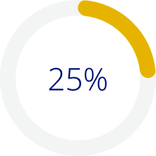
Of people with MASLD
progress to MASH
MASH is a severe progression of unmanaged MASLD. Approximately 25% of people with MASLD are affected by MASH.7
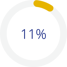
Of people with MASH
may have cirrhosis
Cirrhosis is irreversible scarring/damage to the liver. About 11% of those affected by MASH will develop cirrhosis over several years.7

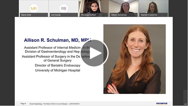
Virtual Medical Expert Training: EUS Liver Evaluation
Participants of the course are instructed on the safe and effective use of Olympus Endoscopic Ultrasound (EUS) equipment through didactics and live-case observations. This program includes advanced EUS applications related to liver disease.
Clinical Support
At Olympus, we strive to be more than just a medical equipment provider to our customers. We provide end-to-end support, from the purchasing process to the procedure and reprocessing services, to build a relationship of trust.
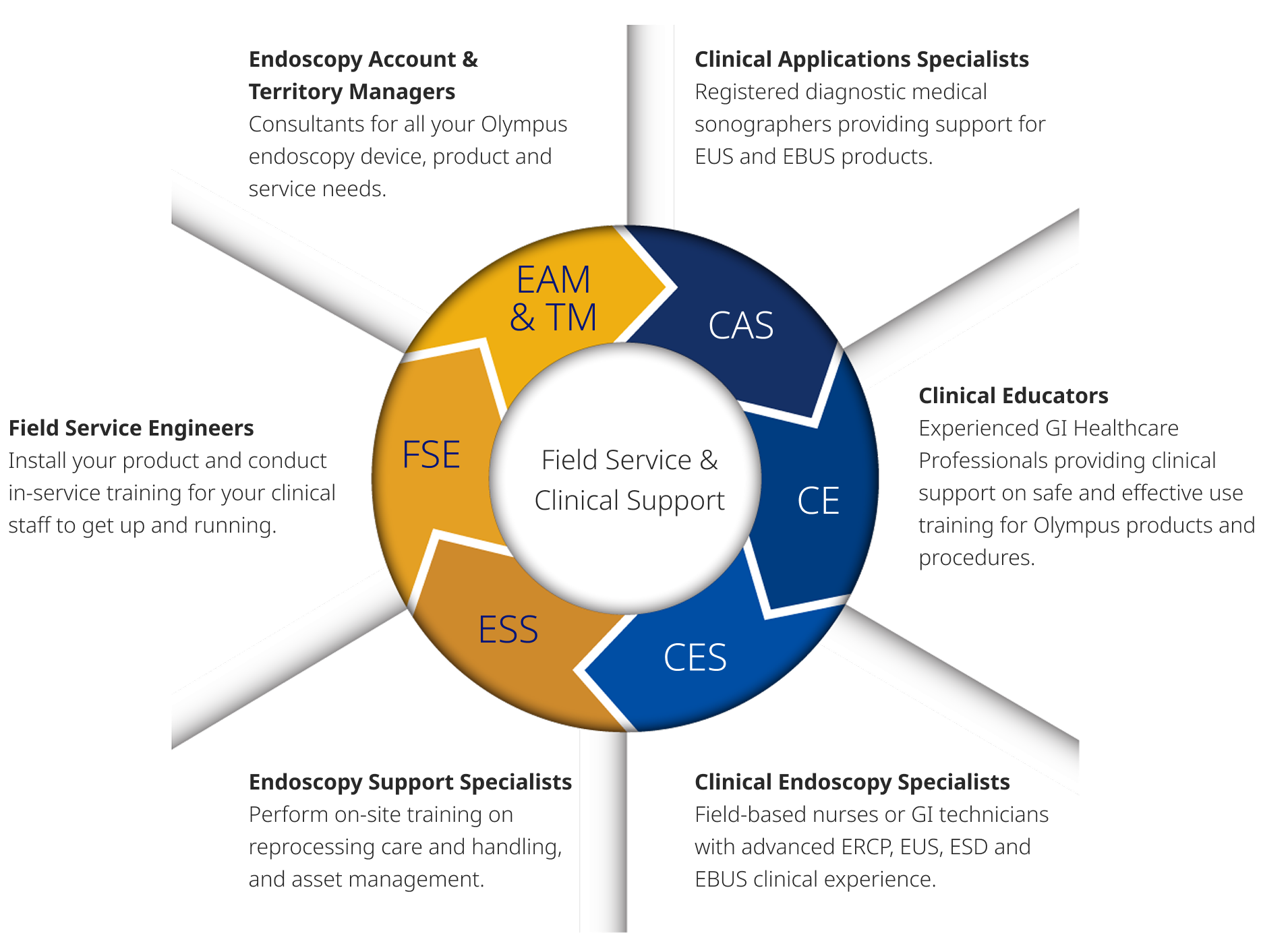
Our Clinical Applications Specialists (CAS) are regionally located, experienced sonographers, dedicated to supporting you and your EUS practice. The CAS team offers on-site and virtual support for evaluations, in-services, installs, training, and procedures. Your CAS will show you how to operate your EUS equipment, offering helpful suggestions for image optimization. Our CAS Team is here to help you achieve success with Olympus EUS products, allowing you to help more patients and reach your goals.
Learn more about your CAS and how they aim to help you treat more patients.
Education
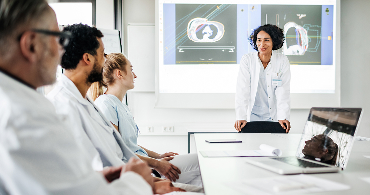
Olympus has a deep commitment to education with a focus on delivering knowledge through science and industry leaders. We pride ourselves in providing exceptional educational support to our customers. We offer a variety of educational opportunities for physicians, nurses, technicians and staff virtually and in-person.
Education
Designed to support you in your pursuit of excellence, Olympus Professional Education is a comprehensive platform of education and training experiences led by healthcare experts from around the world. Learning and training opportunities include hands-on courses, online learning, lectures and workshops, peer-to-peer training, accredited continuing education, and custom on-demand learning as well as a vast library of educational resources.
The Understanding Endoscopic Ultrasound: Technology and Clinical Applications program is designed to train nurses and technicians on the basics of endoscopic ultrasound. The course will cover the physics of ultrasound, GI ultrasound anatomy and ultrasound characteristics and terminology.


Education Library
The Olympus Global Professional Education Library is home to a selection of valuable educational materials including training videos, user guides, quick reference guides, IFUs, channel diagrams and more. The goal of Olympus Professional education is to enhance knowledge, inspire vision and offer solutions to healthcare professionals in the fields of gastroenterology, general surgery, pulmonology, bronchoscopy, urology, gynecology, otolaryngology, bariatrics, orthopedics and anesthesiology.
Diagnostic EUS Training
In collaboration with the American Society for Gastrointestinal Endoscopy (ASGE), our Diagnostic EUS Training course is focused on helping practicing endoscopists based in community hospitals achieve competency in Diagnostic EUS. The program’s online and hands-on curriculum covers the full spectrum of diagnostic EUS and FNA in four to six months in preparation for a proctorship with an EUS expert.
Educational Webinars
Resources
You depend on Olympus to deliver the tools, services, and information you need to provide the highest levels of quality and safety in patient care. To make sure that you have access to advances in endoscopic ultrasound, and remain empowered to make the most of them, we offer education and training programs, comprehensive service and repair programs, and a wide-ranging support network.
White Paper: Endoscopic Ultrasound Fine Needle Biopsy (EUS-FNB) Helps Diagnose Neuroendocrine Tumor of the Pancreas
White Paper: Endoscopic Ultrasound Fine Needle Biopsy (EUS-FNB) Helps Diagnose Gastrointestinal Stromal Tumor (GIST) of Stomach
Publication: Use of the EZ Shot 3 Plus in Endoscopic Ultrasound-Guided Biopsy Procedures
White Paper: How to Optimize EUS Images
White Paper: Current Measurement Parameters for EUS
Watch videos with instructions on how to operate and clean your endoscopic ultrasound echoendoscopes, processors and devices.
Visit Olympus’ pancreatic cancer website to help educate your patients and raise awareness on the importance of early detection. If your facility would like to be included in our Find a Facility resource tool, please contact EUS@olympus.com.
Information on Olympus® OEM service of equipment and access to the Service Portal.
OlympusConnect.com houses the full library of training support, including: Reprocessing Videos, Visual Reprocessing Guides, Instruction Manuals, Reprocessing Manuals, In-service Guides and more.
Sign-up today to receive the latest news and information from Olympus about endoscopic ultrasound (EUS).
For additional questions on service, technical assistance, professional education, or sales contact us directly.
- Data on file with Olympus as if June 8, 2018.
- Data on file with Olympus as of October 31, 2018.
- Data on file with Olympus as of July 27, 2015.
- Data on file with Olympus as of November 16, 1998.
- https://pubmed.ncbi.nlm.nih.gov/32061595/ Accessed September 2025.
- https://gi.org/topics/steatotic-liver-disease-masld/ Accessed September 2025.
- https://thinkliverthinklife.org/wp-content/uploads/2025/07/2025-SLD-Infographic_English.pdf Accessed September 2025.


