


")


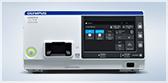
ALOKA ARIETTA 850


Endoscopic Ultrasound
ALOKA ARIETTA 850
Key Benefits
Pure Image: Evolved to enhance your sight
eFocusing
Powered by the ARIETTA 850's advanced Variable Beamformer, eFocusing dynamically performs focusing throughout the depth of field. This transmission and reception technology significantly improves signal to noise ratio (the ratio of intensity between the received signal and background noise), eliminates the need to manually adjust focal zones, and achieves outstanding image clarity from near to far field with less patient-dependent variability.
Active Backend
Active Backend is the powerful image processing engine developed to realize fast, complex arithmetic computations, providing imaging with outstanding definition. New post processing features include:
- Low Echo Reduction: Reduces noise in low echo areas, leaving the brightest areas unchanged.
- Echo Enhancement: Enhances the contrast of the entire ultrasound image by reducing noise and enhancing echoes in the brightest and darkest areas.
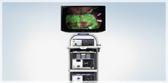
OLED Monitor
The ARIETTA 850 comes equipped with a 22 inch wide OLED (Organic Light Emitting Diode) Monitor for an optimum image display. Without requiring backlighting to function, the OLED Monitor displays true black, allowing a previously unattainable contrast resolution to be achieved. It is the ideal monitor choice for ultrasound, producing the highest quality grayscale display.
Tissue Harmonic Imaging (THI)
When ultrasound waves are propagated through tissue, distortion occurs and harmonic components are generated. THI uses these components to build an image of the targeted area. Advantages of harmonic imaging include improved resolution, improved signal-to-noise ratio, and reduced artifacts. The ARIETTA 850 comes equipped with High Definition (HdT) Tissue Harmonic Imaging. HdT mode with the GF-UCT180 scope has comparable sensitivity to B mode imaging on the ProSound F75 at 7.5MHz, but with a higher resolution.
Advanced Technology: Evolved to elevate your examination
Real-time Tissue Elastography (RTE)
Optional software package for the ARIETTA 850. Included with the ARIETTA 850 AT.
RTE is an additional tool for use in tissue diagnosis. This imaging modality assesses tissue strain in real time and displays the measured differences in tissue stiffness as a color map. The color image shows tissue elasticity, or stiffness in the area of interest, which may help indicate disease processes. This may help the physician identify the appropriate areas for biopsy.
Displacement Direction Guide
Included with RTE Software
RTE now comes equipped with a Displacement Direction Guide, which displays, in real time, the direction of tissue displacement with respect to the probe surface. This guide can help the physician construct highly accurate elasticity images, when using it to check the direction of tissue displacement.
Auto Frame Selection: Select Frame
Included with RTE Software
The best frame for elasticity evaluation can be selected automatically by using the Select Frame function. Select Frame works on the basis of analyzing the stability of the curve on a strain graph and the amount of sampling data.
Strain Ratio
Included with RTE Software
RTE also has the capability of calculating a strain ratio measurement. Strain ratio allows the user to specify two regions of interest (ROI) on the RTE image, one containing normal tissue and one containing abnormal tissue. The system will then analyze the flexure within the ROIs and calculate a quantitative output, relating the elasticity of the two ROIs to one another.
Assist Strain Ratio
Included with RTE Software
The system has the ability to automatically select the ideal locations for the strain ratio ROIs. Using this feature in combination with Select Frame, could potentially reduce the amount of time required to obtain a strain ratio measurement.
Strain Histogram Measurement
Included with RTE Software
The Strain Histogram feature on the ARIETTA 850, displays the hue histogram for the values of relative strain within a region of interest. A hue histogram is a representation of the distribution of colors in an image. This feature calculates the quantitative characteristics of the elasticity image.
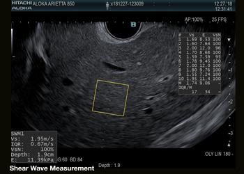
Shear Wave Measurement (SWM)
Optional software package for the ARIETTA 850. Included with the ARIETTA 850 AT.
SWM is used to assess tissue stiffness, such as liver fibrosis. A “push-pulse” generates shear waves, which propagate through the tissue, perpendicular to the ultrasound beam. SWM then provides an assessment of tissue stiffness by calculating the propagation velocity of the shear waves (Vs). Hitachi's SWM technology provides an additional reliability indicator (VsN), as an objective evaluation of the Vs measurement. The reliability indicator gives physicians confidence that they are basing clinical decisions on stable and accurate measurement result.
Detective Flow Imaging (DFI)
Optional software package for the ARIETTA 850. Included with the ARIETTA 850 AT.
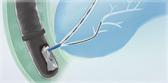
DFI is the new imaging technology for visualization of low velocity blood flow below the previous detection threshold. The unique algorithm displays fine blood flow with greater resolution and sensitivity.
Related Products

Product Support
| Compatible Equipment | |
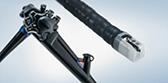
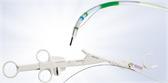
| Electronic Scanning Ultrasound Endoscopes |
GF-UCT180 GF-UE160-AL5 GF-UC140P-AL5 GF-UCT140-AL5 TGF-UC180J BF-UC180F |
| Extracorporeal Probes |
Yes* *Must be purchased through Hitachi Healthcare |
| Ultrasound Functions | |
| Frequency (MHz) | Electronic Scanning: 5, 6, 7.5, 10* *BF-UC180F Scope Only: 5, 7.5, 10, 12 |
| Measurement |
Distance Area Circumference Volume Velocity Flow Volume Strain Ratio Strain Histogram Shear Wave |
| Viewing monitor | |
| OLED Monitor | 22.0 inch, 16:9 aspect ratio |
| Monitor Movement |
Rotation: 160° Tilt: Upwards 60°, downwards 10° Up and down movement: 224 mm Longitudinal: 225 mm |
| Output | |
| Video Outputs |
DVI-D Y/C Composite SDI* *With included DVI/SDI converter |
| External Storage Device |
CD-R DVD-RAM USB |
| Network | LAN |
| Image Management | |
| Still Image Storage | Yes |
| Movie Clip Storage | Yes |
| Cine Memory | Yes |
| DICOM | Yes, includes wireless DICOM |
| Dimensions | |
| Size (mm) | 550(W) x 900(D) x 1,220-1,695(H) |
| Weight (kg) | 145 |
| Power Supply Conditions | |
| Power Supply Voltage | 100 V to 120 V |
| Electrical Frequency | 50/60Hz |
| Power Consumption | 1300 VA or less |
Olympus Training & Proper Use
Olympus Continuum, is a comprehensive platform of education and training experiences led by healthcare experts from around the world. Learning opportunities include hands-on courses, online learning, lectures and workshops, peer-to-peer training, accredited continuing education, and on-demand learning.
For more information: Olympus Continuum Video