


")


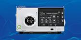
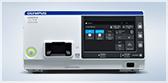
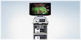
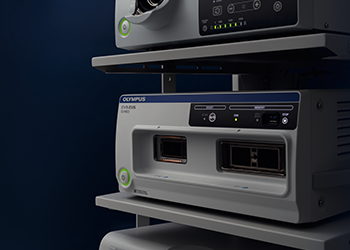
EU-ME3 Ultrasound Processor


Endoscopic Ultrasound
EU-ME3 Ultrasound Processor
Key Benefits
Advancing the Dimensions of Endosonography
Focused on Your Expertise
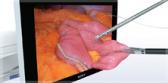
- B Mode - The EU-ME3 provides high-resolution imaging, bringing real clarity to your EUS and EBUS procedures to support detection and characterization of lesions.1
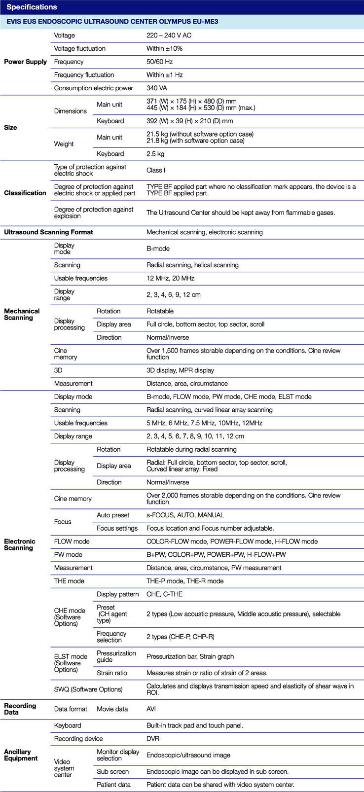
- s-FOCUS - The EU-ME3 is equipped with an s-FOCUS mode that performs focusing throughout the depth of field, eliminating the need to make manual adjustments of the focal zones during the procedure.2
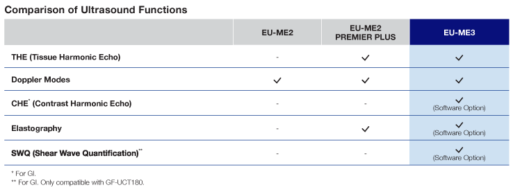
- Tissue Harmonic Echo (THE) - When ultrasound waves are propagated through tissue, distortion occurs, and higher-frequency harmonic components are generated. THE uses these components to build an image of the targeted area, providing a detailed depiction. Advantages of harmonic imaging include improved resolution, improved signal-to-noise ratio, and fewer artifacts.3
- Doppler Modes - The EU-ME3 offers four Doppler modes to distinguish blood flow - Color Flow, Power Flow, Pulsed Wave Doppler (PWD) and H-Flow. H-Flow is a sensitive doppler that shows directional blood flow with less blooming and is especially useful for imaging small vessels. Doppler modes can be used to support safe procedures, which can be a benefit to both the patient and the physician.4
- Contrast Harmonic Echo (CHE) - CHE visualizes harmonic components from ultrasound contrast agents to observe blood flow or microvascularity within tissues. C-THE mode derives images from signals produced by the tissue and the contrast agent, simultaneously.5
- Shear Wave Quantification (SWQ) - SWQ provides an absolute value of tissue stiffness within a region of interest. It performs a quantitative tissue assessment by calculating the propagation velocity of shear waves, generated from a push-pulse.6
- Elastography (ELST) - The EU-ME3 features elastography, which visualizes the amount of relative strain in the tissue (tissue stiffness) during compression and retraction, making it possible to obtain more information about tissue properties. i-ELST is a technology incorporated into the elastography mode that makes it easy to display stiffness color maps, even when tissue displacement is modest.7
- Keyboard Usability - The keyboard was designed with a simple layout in mind and includes a user-friendly built-in touch panel, LED backlit keys and a trackpad for ease of use and cleaning. The large LCD touch panel allows for a wide range of functions to be displayed at one time.
- Programmable User Settings - Users can customize and save imaging parameters as presets, so examinations can always be conducted with the desired settings.
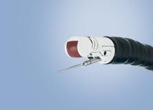
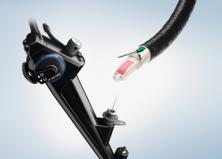
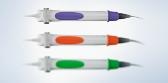
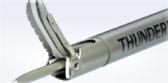
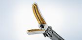
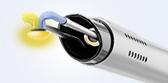
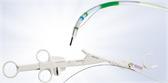
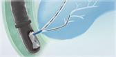
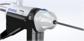
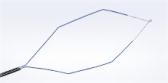
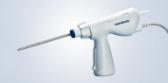
- Wide Range of Compatibility - Integrating both electronic and mechanical scanning technologies, the EU-ME3 is compatible with Olympus® echoendoscopes and miniature probes, creating a total endosonography solution for a full range of applications.
- Customizable Features - Software options are available to meet the needs of your facility. Optional functions allow you to customize your system according to your needs and budget, at any time.
EUS Processor Risks:
High output and prolonged exposure to ultrasonic waves can adversely affect the internal tissues of the patient. Scan only for the minimum length of time necessary for the diagnosis, and at the lowest possible output. Improper care, installation, or use, can cause electric shocks, burns or other injuries. The ultrasound endoscope connected to this ultrasound center must never be applied directly to the heart as it could cause ventricular fibrillation or otherwise seriously affect the cardiac function of the patient. Never allow an EndoTherapy accessory or another ultrasound endoscope, applied to or near the heart, to come in contact with the ultrasound endoscope connected to this ultrasound center. Do not use contrast agents when using the shear wave function, as there is a risk of injury to the patient’s tissue, resulting in bleeding, due to the interaction between the acoustic pressure of the ultrasound and the contrast agent. Do not use the shear wave function during puncturing or the interposition of any other type of metal as it may cause problems during the procedure. Contrast imaging and shear wave elastography functions are for GI only.
Images are from the EU-ME3 with the GF-UCT180 scope, provided by Prof. Dr. Pietro Fusaroli - University of Bologna at Hospital of Imola, Italy and Dr. Khanh Do-Cong Pham - Haukeland University Hospital, Norway
- Data on file with Olympus (DC00580990, DC00849186)
- Data on file with Olympus (DC00580990)
- Data on file with Olympus (DC00626608)
- Data on file with Olympus (DC00567894)
- Data on file with Olympus (DC00626608)
- Data on file with Olympus (DC00580990)
- Data on file with Olympus (DC00580990, DC00623250)
Product Support
Olympus® Service & Repair
Olympus offers a broad range of services to healthcare professionals and to our customers, including contact hour and peer-based training courses; information, training tools and videos on infection control and reprocessing; authorized repair services and support on Olympus equipment; and financing solutions to help your facility with acquisition of new capital equipment, accessories, and maintenance plans.
Need Help?
Cleaning, Disinfection & Sterilization
The proper cleaning, disinfection, and sterilization of Olympus equipment is equally as important as their proper use.
| View Products | Information | Instructional Videos | Customer Portal |
|---|---|---|---|
| OER-Elite | Infection Prevention | GI Endoscope Reprocessing | Log-in |
| All Products | Alerts & Statements | Ultrasound Reprocessing | Register |
| Resources |
Olympus Training & Proper Use
Olympus Continuum™, is a comprehensive platform of education and training experiences led by healthcare experts from around the world. Learning opportunities include hands-on courses, online learning, lectures and workshops, peer-to-peer training, accredited continuing education, and on-demand learning.
For more information: Olympus Continuum™ Video