


")


Every Adenoma Counts
Deadly, Yet Preventable
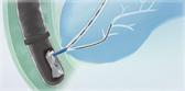
Colorectal cancer (CRC) is one of the most preventable cancers, yet is the second leading cause of cancer-related deaths.1 The key to reducing CRC mortality is encouraging patients to adhere to screening recommendations, and screening colonoscopy is the gold standard.2
Quality Indicators for Colonoscopy
What is Your ADR?
Adenomas are precancerous growths. Adenoma detection rate (ADR) is the number one quality indicator for a colonoscopy. ADR refers to how often an endoscopist finds at least one adenoma during a CRC screening.2

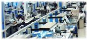
The Big Picture for
Successful Colonoscopy
ASGE/ACG 2024 Colonoscopy Quality Indicators
The American Society for Gastrointestinal Endoscopy (ASGE)/American College of Gastroenterology (ACG) Quality Task Force recommend increasing the target ADR, a priority indicator, from 25% to 35% for endoscopists. A sessile serrated lesion detection rate was introduced with a target of ≥ 6%. The Task Force recommends interventions including the ones highlighted in Olympus’ “Solutions for Successful Colon Cancer Screening” brochure.2,4
Get more data on CRC and ADR!
Download the “Solutions for Successful Colon Cancer Screening” brochure, or contact us to learn more!
Olympus® Solutions for Improving ADR
To help increase ADR, Olympus offers distinct yet complementary solutions: Imaging technology, computer aided detection (CADe) + artificial intelligence (AI) software, mechanically enhanced colonoscopy, and tools to support technique.
Texture and Color Enhancement Imaging (TXI™) Technology
See Things in a New Light
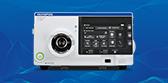
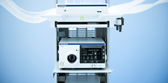
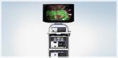
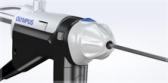
The EVIS X1™ endoscopy system is our most advanced endoscopy solution, which features TXI™ technology, designed to increase the visibility of potentially suspicious lesions and polyps by enhancing imaging color and texture during endoscopic screening.6
TXI Technology — 13.61% ADR Improvement7
In a randomized, controlled trial, TXI technology demonstrated a significant improvement in ADR and the rate of adenomas per colonoscopy ≥ 5 mm in size, versus white light endoscopy (WLE).7
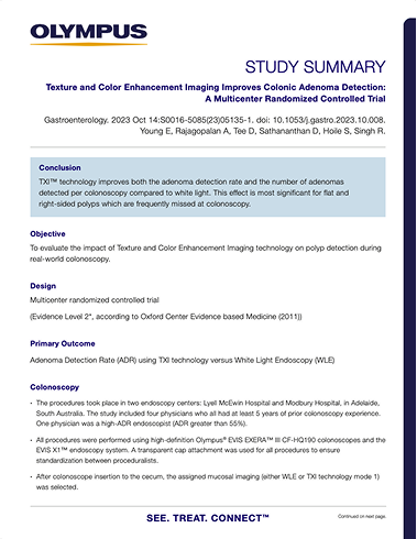
TXI technology is not intended to replace histopathological sampling as a means of diagnosis.
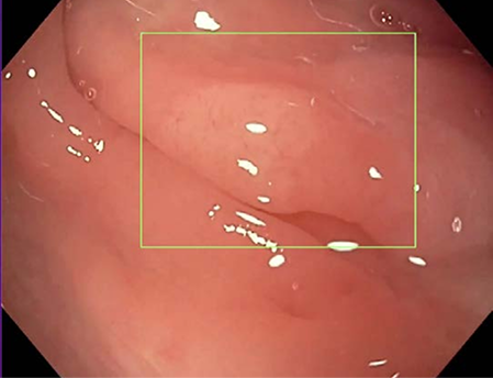
Image captured using CF-H190I scope.
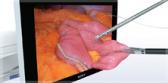
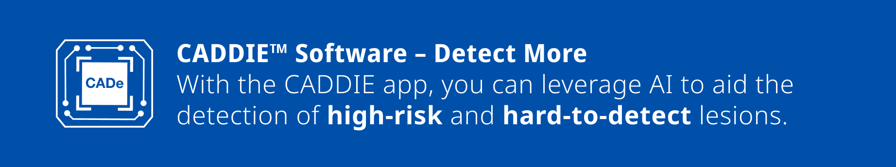
High-Risk and Hard-to-Detect Lesions
Combining human expertise with data driven insights, the CADDIE software aids detection of high-risk lesions—such as sessile serrated lesions (SSLs), large polyps and adenomas—as well as hard-to-detect SSLs and non-polypoid lesions. This capability has generated the following results (per colonoscopy)8:
- 136% relative increase in the detection of large polyps (>10mm).8
- 93% relative increase in the detection of large adenomas (>10mm).8
- 57% relative increase in the detection of flat adenomas.8
- 230% relative increase in the detection of sessile serrated lesions (SSL).8
The gastroenterologist is responsible for reviewing CADDIE suspected polyp areas and confirming the presence or absence of a polyp based on their own medical judgment.
CADDIE is not intended to replace a full patient evaluation, nor is it intended to be relied upon to make a primary interpretation of endoscopic procedures, medical diagnosis, or recommendations of treatment/course of action for patients. The CADDIE computer-assisted detection device is limited for use with standard white-light endoscopy imaging only.
Reach out about the OLYSENSE™ AI platform with CADDIE software!
An Olympus representative would be happy to demonstrate the potential
this technology can unlock.
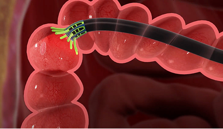
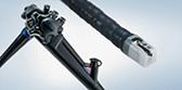
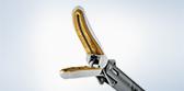
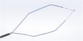
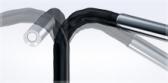
ENDOCUFF Vision™ Mechanically Enhanced Colonoscopy
The ENDOCUFF VISION device attaches to the distal end of a colonoscope and is designed to mechanically enhance mucosal exposure, allowing the endoscopist to manipulate colonic folds. The device is intended to improve visualization and control, which may assist in the detection of abnormalities in the colon.9
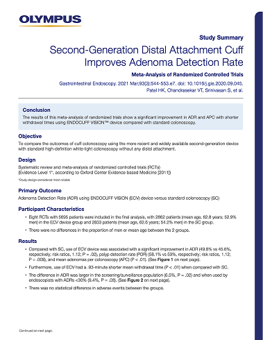
ENDOCUFF Vision – 6.5% to 9.4% ADR Improvement10,22
In a meta-analysis, the ENDOCUFF VISION device resulted in a notable increase of 9.4% in adenoma detection rate (ADR), with range up to 15.1% resulting in higher detection performance amongst operators with low baseline ADR compared to standard colonoscopy.10,22
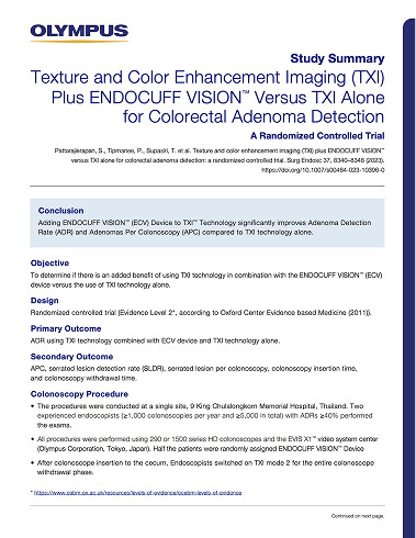
ENDOCUFF Vision + TXI Technology – 13.5% ADR Improvement11
In a prospective randomized controlled trial, ADR was shown to be significantly higher when ECV was used with TXI technology, compared to using TXI technology alone.11
A rare but potential complication when using ENDOCUFF VISION™ device is the detachment of the device during the procedure. Be prepared to retrieve the device if this were to occur. Ensure ENDOCUFF VISION™ device is used only with compatible colonoscopes, that the colonoscope distal tip is in good condition, and that the device is fully seated on the distal tip prior to the application of lubricant to minimize the chance of detachment.
Tools to Support Technique:
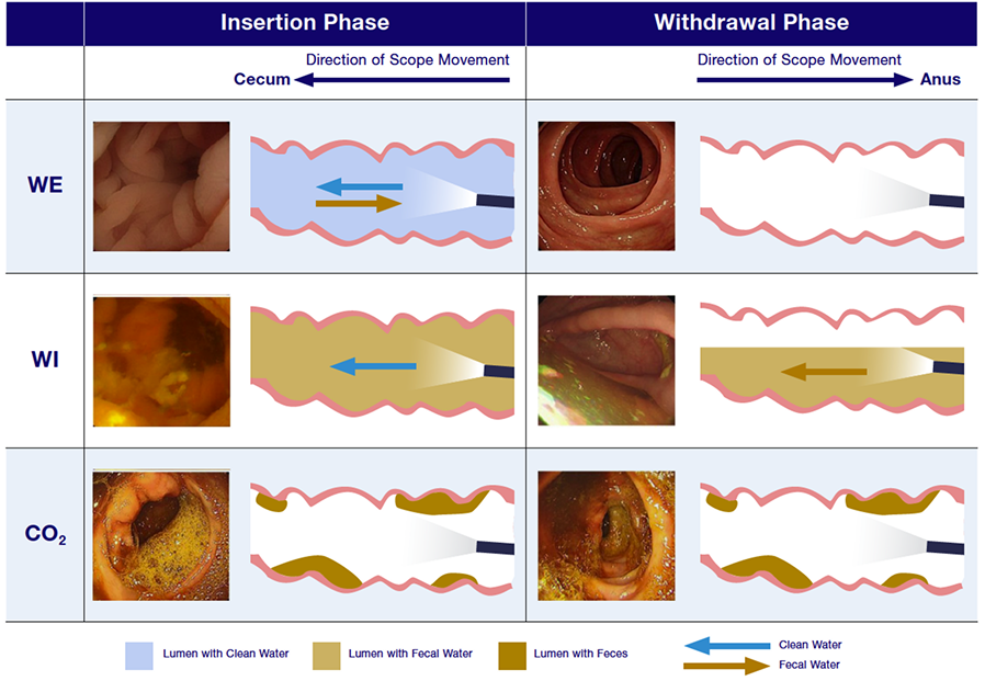
Water-aided Colonoscopy
Water-aided colonoscopy (a.k.a. water-aided endoscopy, or water-based technique), is a practice employed to support scope advancement as an alternative to CO2 or air insufflation, with the added benefit of enhancing bowel cleanliness and providing a magnification effect of the colon.12
Supporting tools include:
- The Olympus® OFP-2 flushing pump, which enables efficient irrigation through endoscopes to improve mucosal visibility23
- The Olympus® KV-6 suction pump, designed for endoscopic aspiration24
Water-aided Colonoscopy – 1.41 x More Likely to Detect Serrated Adenoma12
In a network meta-analysis, colonoscopists were 1.41 times more likely to find at least one serrated adenoma per colonoscopy when water-aided colonoscopy techniques were used, compared to HD WLE alone.12
Dig Deeper into the Olympus®
CRC Screening Portfolio –
How Do These Technologies Work?
Download our comprehensive brochure that outlines the current portfolio and explains how these technologies work. Plus, follow QR coded links to study summaries on the growing body of research that supports the Olympus® CRC Screening portfolio!
Olympus® Solutions for Visualization
Olympus has several technologies to enhance visualization during a colonoscopy.
These interventions include A 4K UHD medical grade monitor, Narrow Band Imaging™ (NBI™) Technology, and Dual Focus technology.
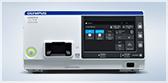
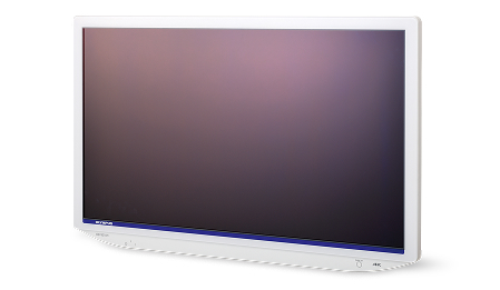
The OEV321UH Display Monitor
Do You See What I See?
The OEV321UH display monitor completes the image chain to provide high-gradation images and a breadth of features designed to facilitate optimal image quality and ease-of-use.13
Narrow Band Imaging™ (NBI™) Technology
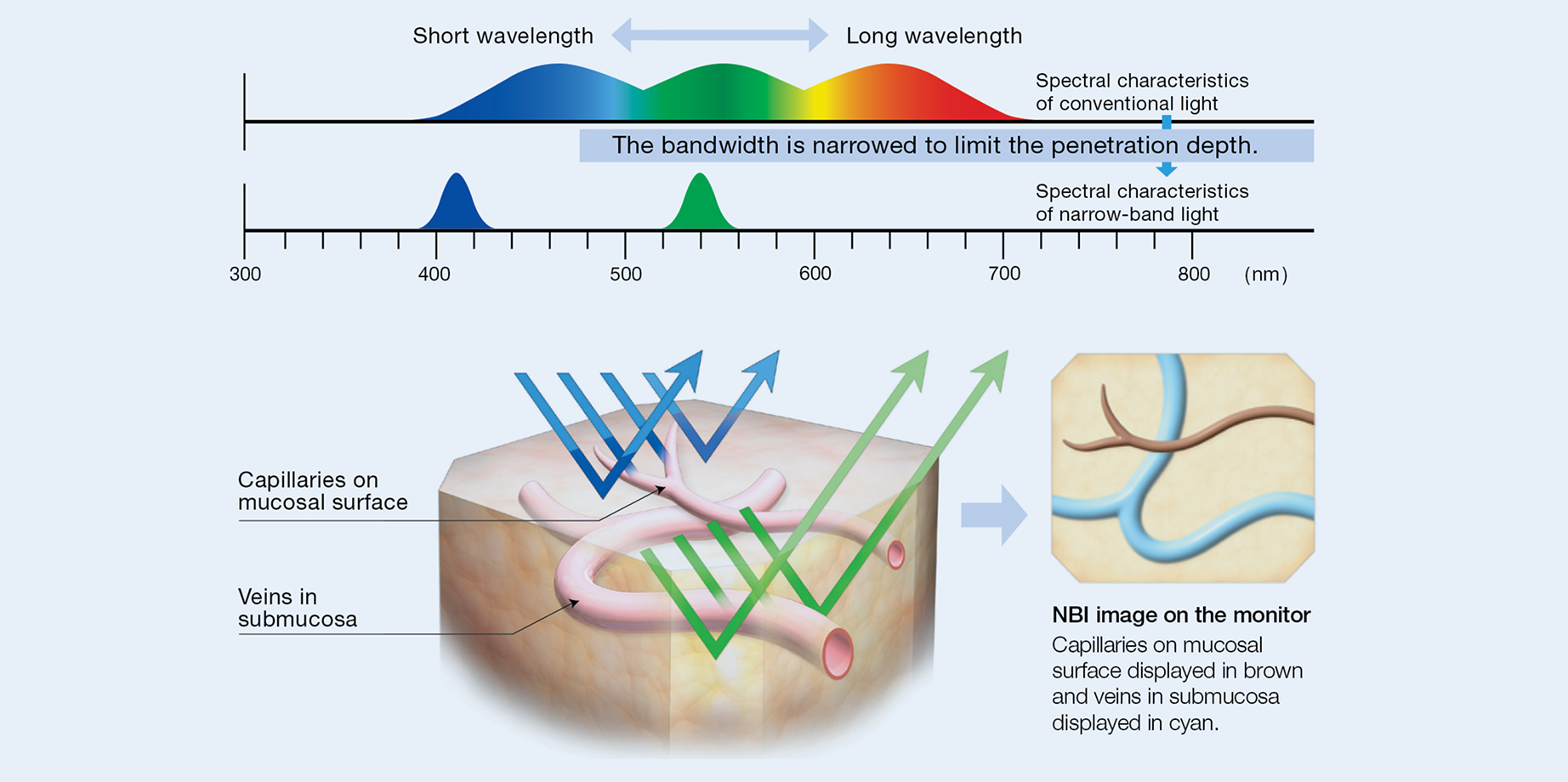
Suspect Before You Resect. NBI™ Technology enhances visualization of the capillary network and mucosal morphology.6 NBI™ Technology assists an experienced endoscopist employing a validated polyp classification system such as the NBI International Colorectal Endoscopic (NICE) classification with high confidence, in distinguishing diminutive adenomatous polyps from non-adenomatous polyps during colonoscopy with greater than 93% sensitivity, 85% specificity and a greater than 90% negative predictive value.14
NBI™ Technology is not intended to replace histopathological sampling as a means of diagnosis.
Dual Focus Technology
Bringing It Into Focus. Dual Focus is a two-stage optical lens technology designed to allow physicians to switch from normal to near focus mode with a single button, to conduct close examination of mucosal tissue and capillary networks. When Dual Focus and NBI™ Technology are used together, endoscopists are more likely to make high confidence predictions of diminutive polyp histology than those using standard focus colonoscopes.15
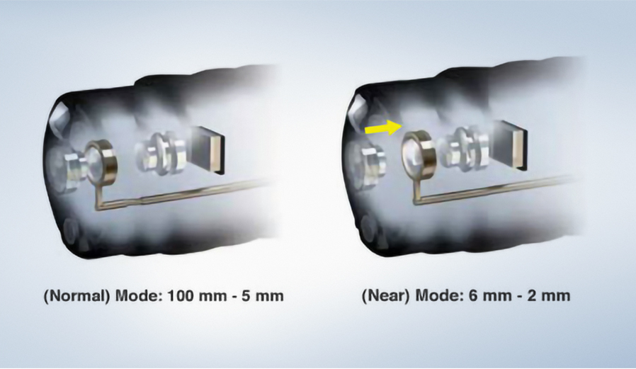
GIF-EZ1500 gastroscopes.
EDOF™ Technoloy
Bring More Into Focus -- Extended Depth of Field technology (EDOF™) Technology
EDOF technology creates an image in total focus by capturing near- and far-focused images simultaneously and combining them into one image with a wide depth of field. It allows the EZ1500 series scopes to provide closer-focused observation in near and normal modes, as well as higher magnification, than the GIF-HQ190 and CF-HQ190L/I scopes.25
Find out more about Olympus® solutions for visualization today!
Download the "Solutions for Successful Colon Cancer Screening" brochure, or contact us to learn more!
Olympus® Solutions for Intubation
Reaching the cecum is the first key step in a colonoscopy.
Olympus offers products to support cecal intubation, such as scope mapping,
specialized colonoscope technologies, and insufflation technology.
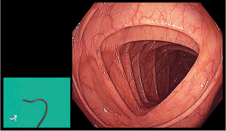
ScopeGuide™ Technology
Recognize loops as they form. ScopeGuide technology is designed to provide a real-time, 3D image of the position and configuration of the colonoscope during a procedure. It is designed to help physicians recognize loops as they form, potentially decreasing insertion time and lessening patient discomfort.16
Responsive Insertion Technology (RIT)
Tactile colonoscope technology in most* of Olympus® 190-, 1100- and EZ1500-series colonoscopes features Responsive Insertion Technology (RIT). RIT combines three proprietary insertion tube technologies:17,18
- High Force Transmission
- Passive Bending
- Variable stiffness technology
*PCF-PH190L/I only has Passive Bending and High Force Transmission
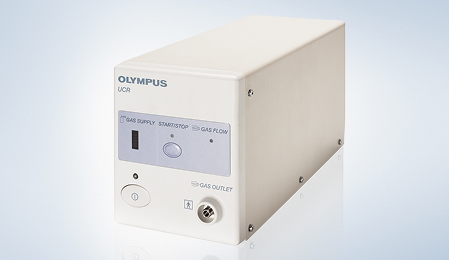
CO2 Insufflator: Advance the Scope by Distending and Expanding the Colon
CO2 Insufflators like the UCR CO2 regulation unit provides intraluminal insufflation using carbon dioxide to minimize bowel distension.19 Because CO2 is absorbed by the body significantly faster than air, the technology is designed to reduce patient discomfort resulting from bowel distension.
CRC Treatment
Olympus is not just dedicated to technology related to early CRC detection.
We offer snares, biopsy forceps, and EndoTherapy devices designed for the evaluation
and treatment of GI diseases.
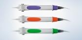
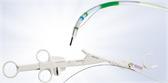
SnareMaster™ Snares for Polypectomy
SnareMaster™ Snares are designed with a thin loop wire facilitates sharp cold cutting ability. These snares come in variety of sizes to customize technique based on need.20
ASGE/ACG Quality Task Force updated 2024 quality indicators and recommended the use of a cold snare technique in ≥ 90% of polypectomy for 4- to 9-mm polyps.2 The SnareMaster Plus snare is ideally suited for this purpose.

EndoJaw™ Biopsy Forceps
Get answers. Olympus offers an extensive range of single-use forceps designed for performance and a range of sizes to optimize the task at hand.21
Do not use these instruments for any purpose other than their intended use as this could cause patient injury or damage to the instrument. Performing polypectomy within the GI tract is a technically demanding procedure and use of associated products such as the SnareMaster™ Plus electrosurgical snare may result in possible electrosurgical risks and patient injury to include, but are not limited to, infection, bleeding, perforation, and mucous membrane damage. When applied to a patient with a pacemaker implanted, the instrument or a cord may cause malfunctioning or failure of the pacemaker, seriously affecting the patient. Before proceeding, always confirm with a cardiologist or the manufacturer of the pacemaker that it is safe to proceed.
Olympus® Endoscopic Devices
Product Catalog
Download our comprehensive product catalog that covers the entire Olympus® EndoTherapy portfolio, with color coding and interactive features.
Service and Support Centered Around You
Our People are Here to Support Your GI Team
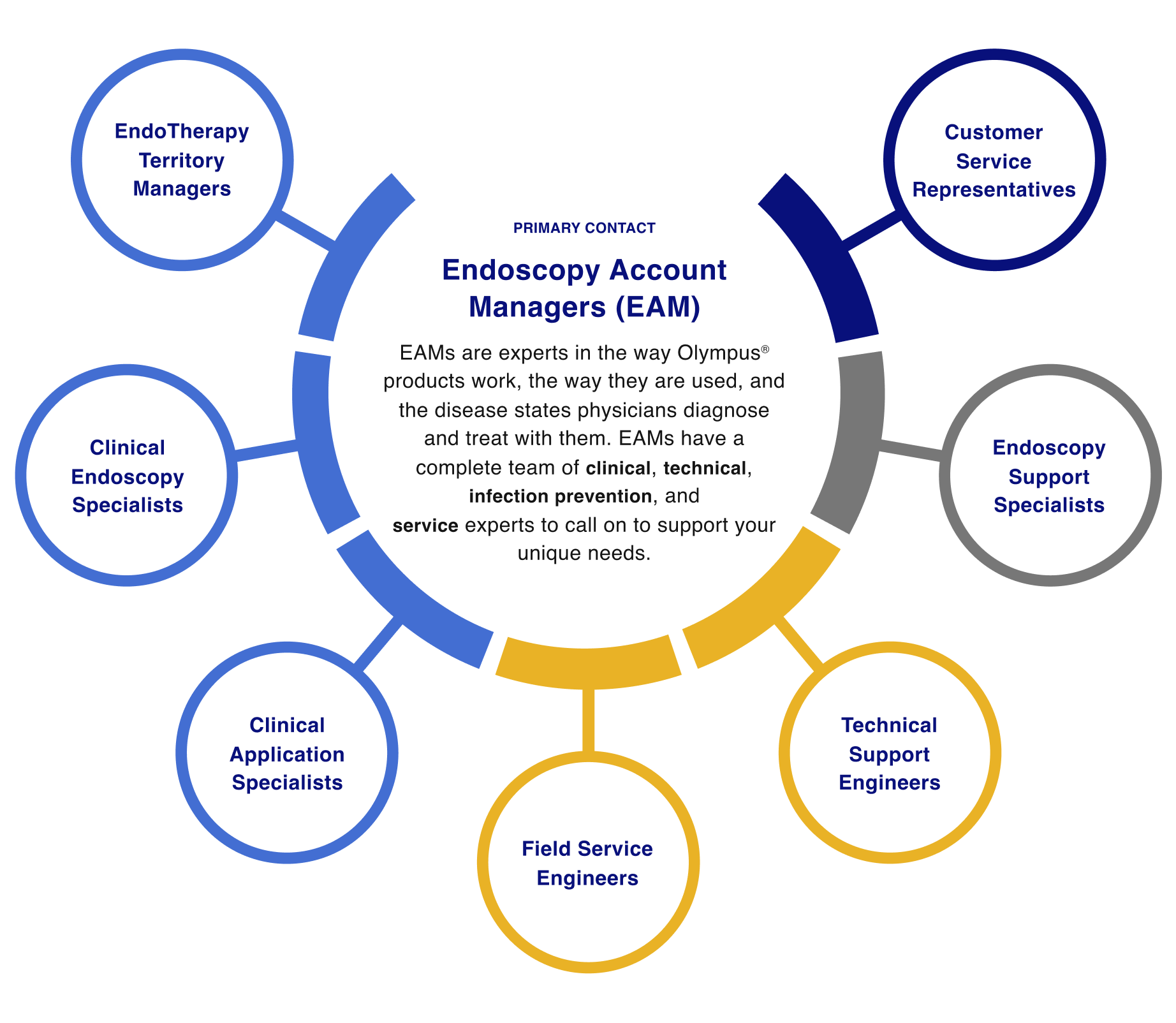
Clinical Support and Education
EndoTherapy Territory Managers (ETM)
ETMs lead ongoing product training sessions and in-services for EndoTherapy products both during procedures and in between cases.
Clinical Endoscopy Specialists (CES)
The CES’s mission is to ensure you are using your equipment to itts fullest potential.
Clinical Application Specialists (CAS)
The CAS are regionally located, registered diagnostic medical sonographers providing support for EUS and EBUS products.
Service
Customer Service Representatives
Calls and requests are handled quickly and efficiently, with an emphasis on keeping you and your team performing at maximum efficiency.
Technical Support
Field Service Engineers (FSE)
The goal of every FSE is to maximize your uptime so you can keep patient care on track.
Technical Support Engineers (TSE)
The TSE’s mission is to keep your equipment running smoothly every day.
Infection Prevention Support
Endoscopy Support Specialists (ESS)
An ESS concentrates their efforts on assisting you to get the most out of your Olympus product's performance.
See how Olympus can support improved outcomes
in your CRC screening program!
Complete the form below for a product demo or to learn more from an Olympus® representative!
- Seer.cancer.gov. Cancer Stat Facts: Common Cancer Sites. Accessed December 9, 2024.
- Rex DK, Anderson JC, Butterly LF, et al. Quality indicators for colonoscopy. Gastrointest Endosc. 2024;100(3):352-381.
- Zhao S, Wang S, Pan P, et al. Magnitude, Risk Factors, and Factors Associated With Adenoma Miss Rate of Tandem Colonoscopy: A Systematic Review and Meta-analysis. Gastroenterology. 2019;156(6):1661-1674.e11.
- ASGE.org. ASGE and ACG Release Updated Quality Indicators for Colonoscopy. Accessed April 22, 2025.
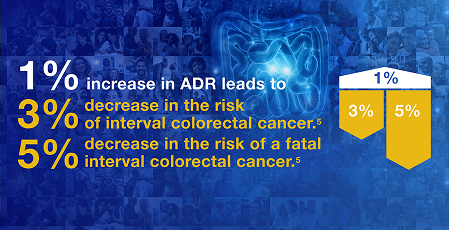
- Corley DA, Jensen CD, Marks AR, et al. Adenoma detection rate and risk of colorectal cancer and death. N Engl J Med. 2014;370(14):1298-1306.
- Data on file with Olympus (DC00489968).
- Young E, Rajagopalan A, Tee D, et al. Texture and Color Enhancement Imaging Improves Colonic Adenoma Detection: A Multicenter Randomized Controlled Trial. Gastroenterology. 2024;166(2):338-340.e3.
- Data not peer-reviewed/published at time of document creation, Data on file with Odin Medical Ltd, EAGLE Trial - NCT05730192.
- Data on file with Olympus 12/09/2016.
- Patel HK, Chandrasekar VT, Srinivasan S, et al. Second-generation distal attachment cuff improves adenoma detection rate: meta-analysis of randomized controlled trials. Gastrointest Endosc. 2021;93(3):544-553.e7.
- Pattarajierapan S, Tipmanee P, Supasiri T, et al. Texture and color enhancement imaging (TXI) plus endocuff vision versus TXI alone for colorectal adenoma detection: a randomized controlled trial. Surg Endosc. 2023;37(11):8340-8348.
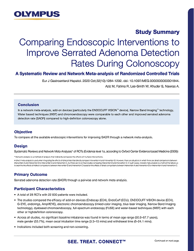
- Aziz M, Fatima R, Lee-Smith W, et al. Comparing endoscopic interventions to improve serrated adenoma detection rates during colonoscopy: a systematic review and network meta-analysis of randomized controlled trials. Eur J Gastroenterol Hepatol. 2020;32(10):1284-1292.
- Data on file with Olympus (DC00438057).
- Data on file with Olympus as of 07/17/2020.
- Kaltenbach T, Rastogi A, Rouse RV, et al. Real-time optical diagnosis for diminutive colorectal polyps using narrow-band imaging: the VALID randomised clinical trial. Gut. 2015;64(10):1569-1577.
- Data on file with Olympus as of 07/Mar/2014.
- Data on file with Olympus 02/16/2012.
- Data on file with Olympus (DC00479164, DC00482967, DC00484835 & DC00364799).
- ASGE Technology Committee, Lo SK, Fujii-Lau LL, et al. The use of carbon dioxide in gastrointestinal endoscopy. Gastrointest Endosc. 2016;83(5):857-865.
- Data on file with Olympus 12/2017.
- Data on file with Olympus 9/28/2021.
- Zorzi M, Hassan C, Battagello J, Antonelli G, et. al, ItaVision Working Group. Adenoma detection by Endocuff-assisted versus standard colonoscopy in an organized screening program: the "ItaVision" randomized controlled trial. Endoscopy. 2022 Feb;54(2):138-147.
- Data on file with Olympus 09/13/2010.
- Data on file with Olympus 04/21/2015.
- Data on file with Olympus (DC00493386, DC00433276, DC00510434 and DC00567392).