


")


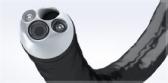
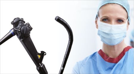
Narrow Band Imaging™ Technology
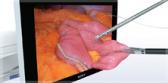
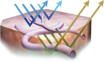
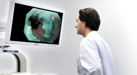
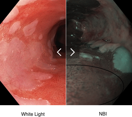
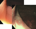
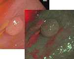
NBI™ Technology is an optical imaging technology that enhances the visibility of vessels and other tissue on the mucosal surface. NBI™ Technology works by filtering the white light into specific light wavelengths that are absorbed by hemoglobin and penetrate only the surface of human tissue. As a result, with Narrow Band Imaging, capillaries on the mucosal surface are displayed in brown and veins in the submucosa are displayed in cyan on the monitor.2
Note: NBI™ technology is not intended to replace histopathology as means of diagnosis
Upper GI Benefits
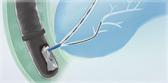
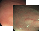
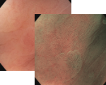
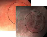
See Before You Sample – High-definition Narrow Band Imaging targets suspicious areas to biopsy for Barrett’s Esophagus.1
Lower GI Benefits
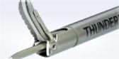
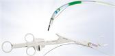
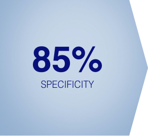
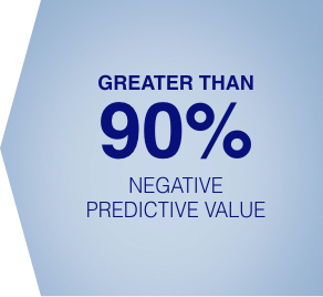
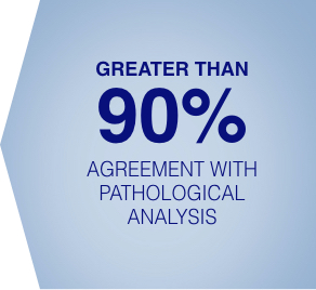
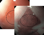
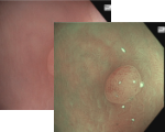
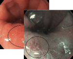
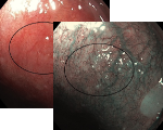
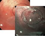
Suspect Before You Resect – High-definition Narrow Band Imaging can be used to assess the histology of diminutive colorectal polyps.2
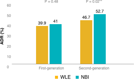
Increased Adenoma Detection Rate – During randomized clinical trials, the use of Second Generation NBI™ Technology resulted in a statistically significant and clinically relevant increase in ADR.5
Suspect Before You Resect


Detection Means Prevention
Increasing adenoma detection rate (ADR) may improve the ability to save lives. A 1% increase in ADR results in a 3% decrease in the risk of interval cancer and a 5% decrease in the risk of a fatal interval colorectal cancer.6
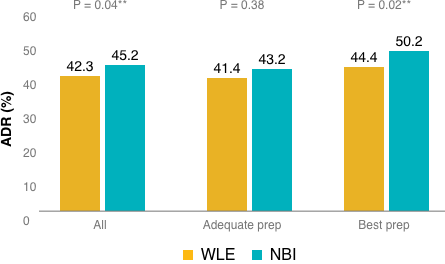
**A p-value less than 0.05 (typically ≤ 0.05) is statistically significant.
No significant difference was observed in the adequate bowel preparation group (OR, 1.07; 95% CI, 0.92–1.24; P = 0.38). However, the odds of detecting at least 1 adenoma in the “best” bowel preparation group was significantly higher with NBI™ Technology compared to WLE (OR, 1.30; 95% CI, 1.04–1.62; P = 0.02**).
(Figure 1)
The odds of detecting at least 1 adenoma with second-generation bright NBI™ Technology* vs white light was significantly higher than with WLE (OR, 1.28; 95% CI, 1.05–1.56; P =0.02); however, this effect was not observed for first-generation NBI™ Technology (OR, 1.06;95% CI, 0.91–1.24; P=0.48).
(Figure 2)
Don't leave biopsy to chance.
Better Interpretation Tool
HD NBI™ Technology provides contrast, which may aid in the interpretation of mucosal morphology, vascular patterns, and blood vessel appearance in patients with Barrett’s esophagus.1
Less Time
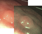
HD NBI™ Technology facilitates targeted biopsies in patients with Barrett’s esophagus, which can save valuable procedure time.1
Fewer Biopsies
Using HD NBI™ Technology to target suspicious areas in Barrett’s.1
Download our GI NBI™ Technology Atlas

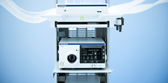
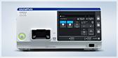
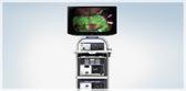
Olympus Continuum™
LEARN MORE


1. Data held on file with Olympus (K100584) as of 07/02/2010
2. Data held on file with Olympus (K192793) as of 07/17/2020
3. ASGE Technology Committee et al. “ASGE Technology Committee systematic review and meta-analysis assessing the ASGE Preservation and Incorporation of Valuable Endoscopic Innovations thresholds for adopting real-time imaging-assisted endoscopic targeted biopsy during endoscopic surveillance of Barrett’s esophagus.” Gastrointestinal endoscopy vol. 83,4 (2016): 684-98.e7. doi:10.1016/j.gie.2016.01.007
4. ASGE Technology Committee et al. “ASGE Technology Committee systematic review and meta-analysis assessing the ASGE PIVI thresholds for adopting real-time endoscopic assessment of the histology of diminutive colorectal polyps.” Gastrointestinal endoscopy vol. 81,3 (2015): 502.e1-502.e16. doi:10.1016/j.gie.2014.12.022
5. Atkinson NSS, Ket S, Bassett P, et al. Gastroenterology. 2019;157:462–71.
6. Corley DA, Jensen CD, Marks AR, et al. Adenoma Detection Rate and Risk of Colorectal Cancer and Death. N Engl J Med. 2014;370(14):1298-1306.
7. Data held on file with Olympus (K121959) as of 01/10/2013
* first-generation NBI™ Technology refers to EVIS LUCERA SPECTRUM™ ENDOSCOPY SYSTEM or EXERA II™ ENDOSCOPY SYSTEM
* second generation NBI™ Technology (bright) refers to EVIS LUCERA ELITE™ ENDOSCOPY SYSTEM or EVIS EXERA III™ ENDOSCOPY SYSTEM
Note: NBI™ Technology is not intended to replace histopathological sampling as a means of diagnosis.