


")


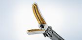
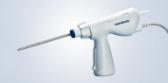
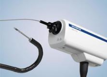
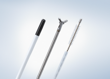
Radial EBUS Probes
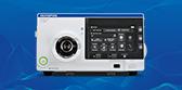
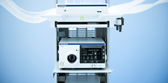
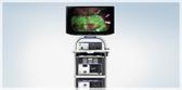
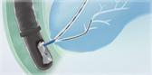
Endobronchial Ultrasound
Radial EBUS Probes
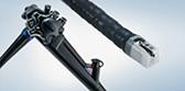
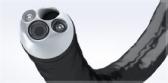
Compatible with a channel diameter of 1.7 mm or more, depending on the model chosen. (See available models below) Radial EBUS probes can be inserted directly through the bronchoscope or used with the Olympus Guide Sheath Kit for an effective solution that can aid in accessing and sampling peripheral pulmonary lesions.
Key Benefits
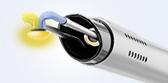
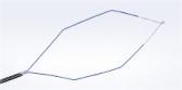
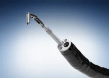
- Precise, real time confirmation: Olympus radial EBUS probes provide real-time confirmation of lesion location.1
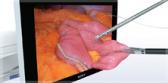
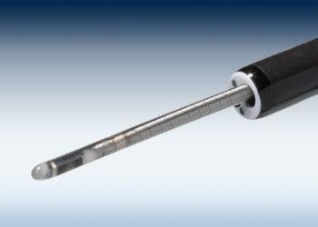
- 360-degree, real time visualization: Radial EBUS provides a 360-degree image of the airway wall and surrounding structures external to the airway. The mechanical scanning technique produces a real-time ultrasound image enabling direct visualization of the lesion location for sampling.1
- Proven technology: Clinical data demonstrates that radial EBUS provides an adequate alternative to conventional transbronchial bronchoscopy, without the increased risk of clinical complications associated with transthoracic needle aspiration (TTNA).2
1. Chen A, Chenna P, Loiselle A, Massoni J, Mayse M, Misselhorn D. Radial probe endobronchial ultrasound for peripheral pulmonary lesions. A 5-year institutional experience. Ann Am Thorac Soc. 2014 May;11(4):578-82. doi: 10.1513/AnnalsATS.201311-384OC. PMID: 24635641.
2. Wang Memoli JS, Nietert PJ, Silvestri GA. Meta-analysis of guided bronchoscopy for the evaluation of the pulmonary nodule. Chest. 2012 Aug;142(2):385-393. doi: 10.1378/chest.11-1764. PMID: 21980059; PMCID: PMC3425336.
Complications from bronchoscopy are rare and most often minor, but if they occur, may include breathing difficulty, vocal cord spasm, hoarseness, slight fever, vomiting, dizziness, bronchial spasm, infection, low blood oxygen, bleeding from the biopsied site, or an allergic reaction to medications. Only rarely do patients experience other more serious complications (for example, collapsed lung, respiratory failure, heart attack and/or cardiac arrhythmia).
Related Products
Product Support
| Item Number | UM-S20-17S |
| Display mode | B-Mode |
| Scanning method | mechanical radial scanning |
| Scanning direction | 360 degree cirumferential, in perpendicular direction with respect to probe insertion direction |
| Ultrasonic frequency (MHz) | 20 |
| Working length (cm) | 215 |
| Total length (cm) | 222.5 |
| Insertion tube (mm) | 1.4 (distal end length 1085) |
| 1.7 (on connector side) |
|
| Compatible scope (mm) | 1.7 or more |
| Contact method for ROI | direct contact method |
| Guide Sheath compatibility | K-401, K-402 |
Olympus® Service & Repair
Olympus offers a broad range of services to healthcare professionals and to our customers, including contact hour and peer-based training courses; information, training tools and videos on infection control and reprocessing; authorized repair services and support on Olympus equipment; and financing solutions to help your facility with acquisition of new capital equipment, accessories, and maintenance plans.
Need Help?
Cleaning, Disinfection & Sterilization
The proper cleaning, disinfection, and sterilization of Olympus equipment is equally as important as their proper use.
| View Products | Information | Instructional Videos | Customer Portal |
|---|---|---|---|
| OER-Elite | Infection Prevention | GI Endoscope Reprocessing | Log-in |
| All Products | Alerts & Statements | Ultrasound Reprocessing | Register |
| Resources |
Olympus Training & Proper Use
Olympus Continuum™, is a comprehensive platform of education and training experiences led by healthcare experts from around the world. Learning opportunities include hands-on courses, online learning, lectures and workshops, peer-to-peer training, accredited continuing education, and on-demand learning.
For more information: Olympus Continuum™ Video